In this article
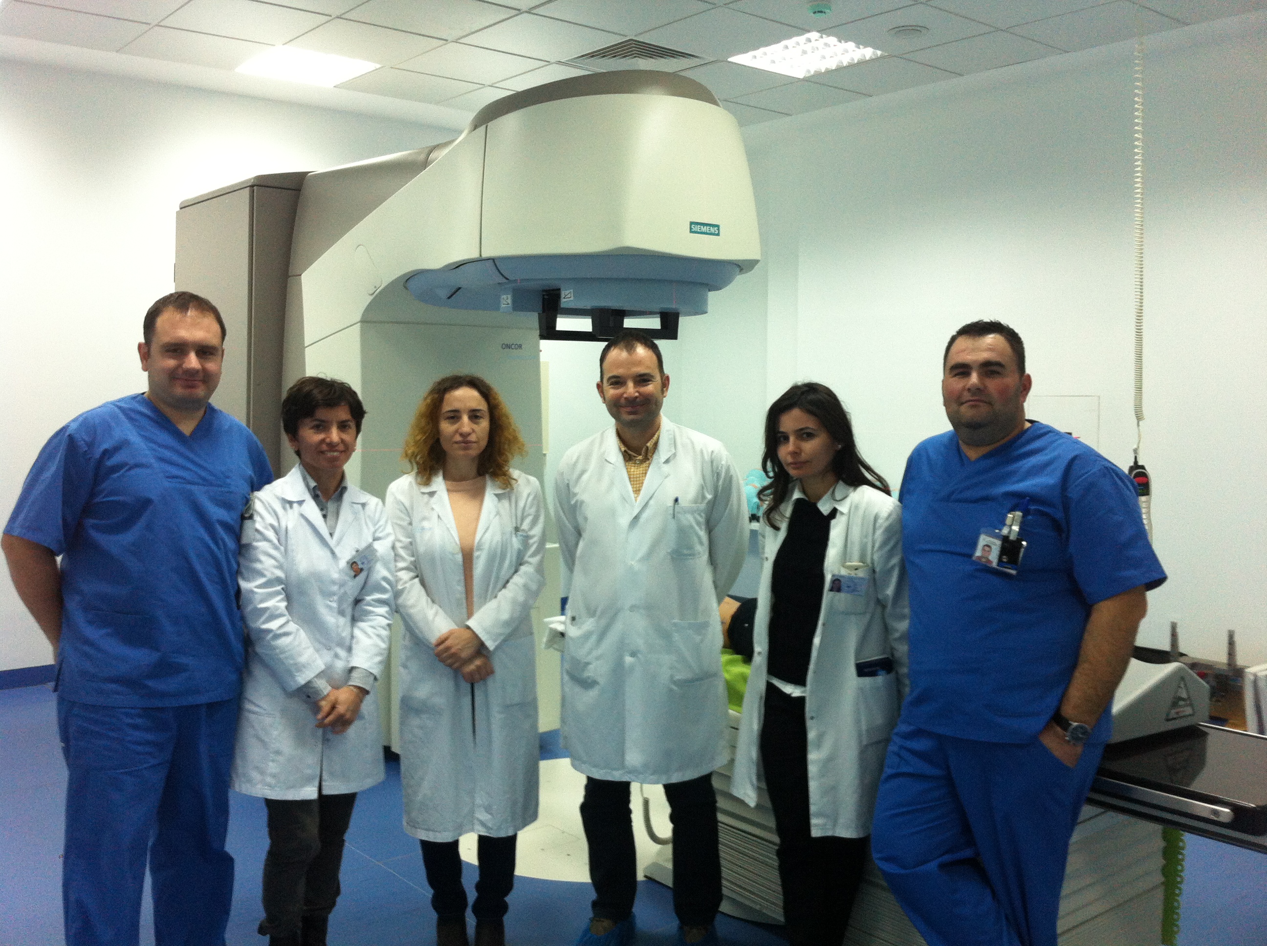 25 tumor cases are treated every day with the X-Knife accelerator at the Professor Kurti Neurosurgery Hospital. Doctors say the machine produces good results but cannot cope with the volume of cases, leaving patients waiting for weeks for treatment.
In an interview with the newspaper Shekulli, neurosurgeon Artur Xhumari says: “The accelerator has made treatment more comfortable and many times more precise for patients with tumors.”
In our country, the fight against tumor diseases is waged with 3 main weapons: surgery, chemotherapy and radiation. The last of these, radiation treatment, has improved significantly this year thanks to the linear accelerator called X-Knife, a modern machine that cost about 1.5 million euros.
Patients treated with radiosurgery no longer have to be admitted to hospital, because their treatment takes place in sessions. In some cases, this also spares them surgical operations, which carry the risk of complications or serious injury.
But although the Mother Teresa University Hospital Center (QSUT) finally has its own accelerator, it is not enough.
“By European standards, one accelerator serves about 500 thousand inhabitants,” says Dr. Artur Xhumari, who adds that at least 6 are needed to meet our country’s needs.
To this end, QSUT’s management is working to have a second accelerator installed during 2015, which would allow more treatments.
 Staff: the key to providing the service
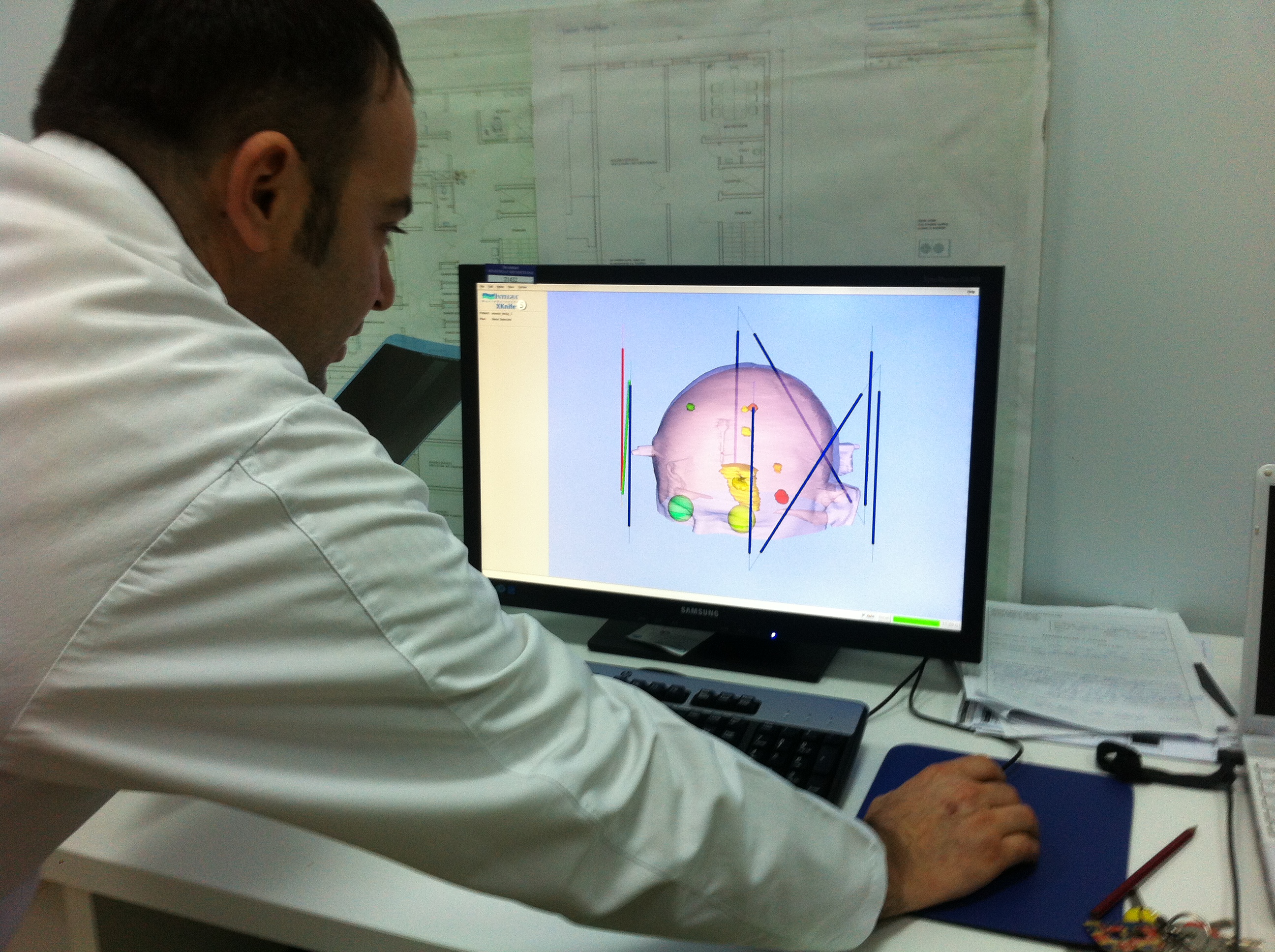
At present, 25 tumor cases a day are treated with the linear accelerator: 24 with radiotherapy and just one with radiosurgery.
According to Dr. Xhumari, the service provided with this machine depends on staff, who must be specially trained.
“The more advanced the equipment, the better prepared the staff must be. Otherwise, the equipment is no use to you,” says Xhumari, adding that the staff is extremely well prepared.
The accelerator team at the Neurology Hospital currently consists of six people: one radiosurgeon (Dr. Artur Xhumari, Doctor of Science), one radiation oncologist (Dr. Aurora Aliraj), two medical physicists (Blerina Myzeqari and Irena Muçollari) and two radiotherapy technicians (Renajdo Tafaj and Bledar Çullhaj).
In this respect, the Neurology Hospital offers something positive: coordination among doctors in treating cases of tumor disease.
“A multidisciplinary team is needed, consisting of the doctor who knows how to operate, then the imaging specialist, and the doctor who understands the disease and treats the patient with medication, who may be a cardiologist, a neurologist and so on. We need the doctor who examines cancer under a microscope, the histopathologist, the biopsy doctor, the anatomical pathologist and others.
In other words, all these specialties are needed to form a multidisciplinary team, which then decides what a patient needs: surgery, chemotherapy or radiation. Here (in neuro-oncology), this multidisciplinary team has existed for years.
“It has now been agreed that other hospitals will also organize such teams, so that other tumor diseases, those that do not affect the brain, can also be managed properly.”
The volume of cases calls for more accelerators
According to Ministry of Health statistics, during 1993-2011 there were an average of about 12,040 admissions a year for tumor diseases in Albania, with a total of 228,759 admissions over this 19-year period.
These numbers are unmanageable even with the new accelerator, which, according to international standards, can cover up to 500 thousand inhabitants.
“We have one accelerator at a time when Albania would need at least six,” says Dr. Xhumari.
But fortunately, according to the doctor, a project is moving forward that envisages adding a second accelerator by the end of next year, until, step by step, there is a third as well.
Yet despite Xhumari’s optimism about future improvement, current conditions mean that people with various tumors have to wait in line for the treatment they need.
The doctor also acknowledges the situation with regret. “A second accelerator would ease the burden enormously, because it is very hard to tell a patient: Yes! You need radiation, but you have to wait your turn, when there is no knowing how long that wait might last.”
 Neurosurgery’s new machine differs from Oncology’s “cobalt” unit

At present, patients at QSUT also receive radiation treatment for tumor diseases at the Oncology Hospital, but that hospital’s machine uses older technology and does not offer the same treatment precision as Neurosurgery’s X-Knife linear accelerator.
“The linear accelerator is used to treat tumors, both malignant and some benign ones. Treatment with this machine is carried out by irradiating tumor cells with photons. Unlike the equipment of earlier years, it does not operate with cobalt, an element that is a radioactive source,” says radiation oncologist Aurora Aliraj.
“Cobalt radiation is a treatment that has been out of use in Europe for about 20-30 years, because the precision of irradiation and dose delivery is lower,” adds radiosurgeon Xhumari.
He goes on to say that the X-Knife accelerator is entirely computerized and has geometric irradiation accuracy of about 1 millimeter.
“This machine also has dose precision. With cobalt, these parameters are less accurate. We must say that, for patients, this difference either fails to provide the required treatment or, in the worst case, causes harm.”
What are tumors?
Tumors are the uncontrolled multiplication of abnormal cells in the human body. They are divided into benign and malignant tumors.
Benign tumors grow slowly, sometimes for years before producing clinical signs. They do not spread to distant sites from the primary site where they formed (in other words, they do not metastasize) and are not infiltrative (there is a clear boundary between the tumor and normal tissue).
Malignant tumors are different. They grow very rapidly, metastasize to distant sites through the blood or lymphatic system, and are infiltrative, meaning there is no clear boundary between the tumor and healthy tissue. This is precisely what makes complete surgical removal of a tumor difficult at the microscopic level.
Symptoms and diagnosis
Dr. Artur Xhumari, Doctor of Science, discusses his own specialty: tumors that affect the brain.
The symptoms of a brain tumor can be general or specific. The latter depend on the tumor’s location, the part in which it appears.
Tumors can present with headaches, epilepsy, balance disorders, neurological deficits (in the form of numbness or paralysis of the limbs), memory disorders and behavioral changes.
There are two circumstances in which a tumor is diagnosed: incidentally (during a routine checkup, when the patient has had no clinical signs up to that point) and as a result of clinical signs appearing.
The diagnosis is based on clinical signs and findings from doctors’ examinations, followed by imaging (a CT scan or MRI; the latter has greater sensitivity in distinguishing a tumor from another brain pathology).
Ultimately, it is the biopsy that confirms the diagnosis.
What causes tumors?
Today, we still do not speak of direct causes of tumors, only of risk factors.
Different tumors have different risk factors. These include smoking, alcohol or drug use, environmental factors, chemicals found in large urban areas, industrial or former industrial areas, ionizing radiation, viruses and so on.
Smoking is considered a risk factor for lung and laryngeal cancer, and Epstein-Barr virus a risk factor for nasopharyngeal cancer and lymphomas.
Genetic predisposition is also considered a risk factor for tumors, and this is clearly seen in breast cancer.
Women who inherit a BRCA-1 gene mutation have a lifetime breast cancer risk ranging from 50 to 85 percent.
Published in “Shekulli” on 5/12/2014
* This article was reviewed and corrected on 8 October 2026.